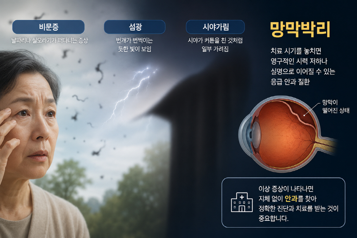
[Sportschosun reporter Jang Jong-ho] Floaters, which can look like tiny flies or threads drifting in front of the eyes, are often seen as a natural part of aging.
However, if floaters are accompanied by flashes of light, like lightning, or if part of your vision is blocked as if a curtain has been drawn, retinal detachment should be suspected. Retinal detachment is an emergency eye disease that can lead to permanent vision loss or blindness if treatment is delayed. If unusual symptoms appear, it is important to visit an eye clinic immediately for an accurate diagnosis and treatment.
◇Retinal detachment, an eye disease that requires emergency treatment
Retinal detachment is a condition in which the retina, located on the inner side of the eye, separates from its normal position. The retina is the tissue that converts light entering the eye into electrical signals and sends them to the brain. When the retina detaches from the inner wall of the eye, its nutrient supply is cut off, the function of the photoreceptor cells gradually declines, and delayed treatment can result in permanent vision damage or blindness.
Professor Jeong Seok-hoon of the Department of Ophthalmology at The Catholic University of Korea Incheon St. Mary's Hospital said, "Retinal detachment is not simply a disease that reduces vision; it is an eye disease that requires emergency treatment." He added, "The earlier treatment begins, especially before the detachment reaches the macula, the greater the chance of preserving vision."
The main cause of retinal detachment is a retinal tear. As the vitreous inside the eye liquefies due to aging or high myopia and separates from the retina, the retina can tear or develop a hole. Fluid then seeps through the opening, causing the retina to detach from the inner wall of the eye. In addition, tractional retinal detachment can occur when fibrous tissue pulls on the retina due to diabetic retinopathy, and exudative retinal detachment can also develop when fluid accumulates because of inflammation.
◇Floaters and flashes of light are the main symptoms... detailed examination is needed
People with high myopia, a family history of the disease, or a history of eye surgery such as cataract surgery are at relatively higher risk of retinal detachment. It can also occur after a strong blow to the eye or other trauma.
The typical symptoms are floaters and photopsia. Floaters are the sensation of black spots, threads, or fly-like shapes drifting in front of the eyes, while photopsia is the perception of flashing light, like lightning, even when no actual light is present. As the disease progresses, one side of the visual field may appear covered by a curtain, objects may look distorted, and vision can drop sharply. If it develops gradually from the peripheral retina, there may be no obvious symptoms, and it may be discovered incidentally during an eye exam.
Professor Jeong explained, "Floaters can also appear as part of the aging process, but if they suddenly increase in number or are accompanied by photopsia or visual field loss, the retina must be checked." He added, "If these symptoms occur, do not delay and undergo a detailed examination."
If retinal detachment is suspected, a dilated fundus examination is performed to inspect the retina after enlarging the pupil. Depending on the case, fundus photography, optical coherence tomography (OCT), and ocular ultrasound may also be used to accurately assess the extent of retinal damage and its condition.
Treatment depends on how much of the retina has detached and whether the macula is involved. If there is only a retinal tear and the detachment is in an early stage, laser photocoagulation can be used to secure the area around the tear and prevent the detachment from progressing. However, if the retina has already detached, surgery is usually required.
Depending on the retinal condition and the extent of detachment, vitrectomy or scleral buckling is performed. Vitrectomy removes the vitreous pulling on the retina, followed by laser treatment, and then gas or silicone oil is injected to help the retina reattach. Scleral buckling is a procedure that uses silicone material on the outside of the eye to press the retina back into its original position. Depending on the patient's retinal condition and the extent of detachment, the two procedures may be performed separately or together. After surgery, if gas has been injected, patients may need to maintain a face-down position for a certain period to improve the treatment effect.
◇If you have high myopia or a family history, regular eye exams are necessary
There is no specific way to prevent retinal detachment, but regular eye exams can help if you are in a high-risk group. If you have high myopia, a family history of retinal disease, or a history of eye surgery, it is advisable to undergo regular fundus examinations even if you have no symptoms. It is also helpful to avoid strong impacts to the eyes and wear protective eyewear during exercise or work if there is a risk of eye trauma.
Professor Jeong emphasized, "Retinal detachment is a disease in which the timing of treatment determines the prognosis for vision." He added, "If floaters suddenly become worse, or if photopsia or blocked vision appears, do not dismiss it as simple fatigue or aging. You should receive an accurate diagnosis and treatment."
Jang Jong-ho, bellho@sportschosun.com