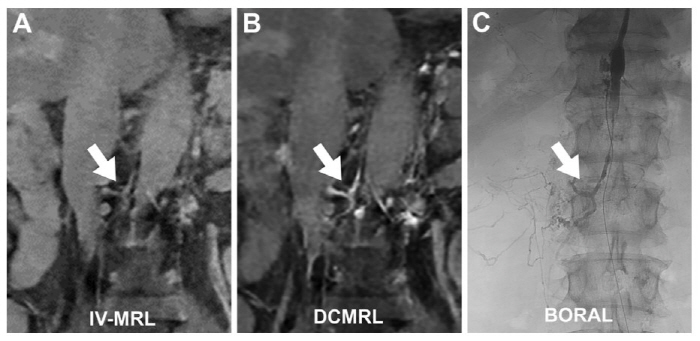
Unlike conventional techniques that require puncturing a lymph node or inserting a catheter (B, C), the lymphatic system can also be easily observed with contrast agent injected through a blood vessel (A). [Sportschosun] A new MRI technique has been developed that can visualize lymphatic vessels using only an intravenous injection, instead of directly injecting contrast agent into the inguinal lymph nodes. Using this method, the team also succeeded in identifying lymphatic vessels in the liver and mesentery, which are difficult to confirm with conventional tests.
The lymphatic system plays an important role in immune function and maintaining fluid balance. However, when lymphatic vessels are damaged during cancer surgery or other procedures, lymphatic leakage can occur, leading to complications such as malnutrition, weakened immunity, and ascites.
For that reason, a test that can quickly and accurately identify the site of leakage is needed. At present, the standard imaging tests for the lymphatic system are inguinal lymph node injection-based lymphangiography and dynamic contrast-enhanced MR lymphangiography (DCMRL).
However, these methods require precise needle placement into the inguinal lymph nodes, making the procedure technically difficult. They also have a major limitation in that they show little of the liver and mesenteric lymphatic vessels, which account for more than 70% of total lymph production.
As a result, it has been difficult to pinpoint the exact location of lymphatic leakage. In particular, for patients with chylous ascites, in which lymphatic fluid accumulates in the abdominal cavity due to mesenteric lymphatic vessel damage, the detection rate for leakage has remained at about 55%, creating diagnostic challenges.
To address this, a research team led by Heo Se-beom of the Department of Radiology at Seoul National University Hospital, Yoon Seong-hwan of the Biomedical Research Institute, and Kwon Ryeo-min of the Department of Radiology at Hallym University Hospital evaluated the effectiveness of a newly developed noninvasive imaging technique, intravenous contrast-enhanced MR lymphangiography (IV-MRL), through animal experiments and a pilot clinical study. First, in a pig model, contrast agent was injected intravenously, and the concentrations in the blood and lymph fluid were compared over time.
The results showed a "lymph phase," in which the contrast concentration in lymph fluid became higher than in blood after 20 to 30 minutes. 5 times compared with the back muscles 20 to 30 minutes after injection, clearly visualizing the lymphatic vessels.
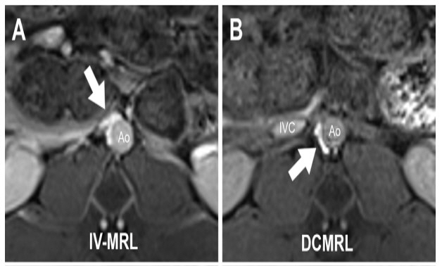
The team also compared intravenous contrast imaging (IV-MRL) with inguinal lymph node injection imaging (DCMRL). Both methods visualized the thoracic duct and retroperitoneal lymphatic vessels, but the intravenous method also visualized the liver and mesenteric lymphatic vessels, which were limited in conventional tests.
Similar results were confirmed in the pilot clinical study. 6 years.
001). These findings show that the new contrast imaging method can identify lymphatic vessels with only an intravenous injection and can also visualize the liver and mesenteric lymphatic vessels that are difficult to detect with existing tests.
Heo Se-beom, professor in the Department of Radiology at Seoul National University Hospital, said, "Unlike conventional lymph node injection methods, IV-MRL can be performed with only an intravenous injection, which can reduce procedural difficulty and the patient's pain burden. " He added, "It is expected to help improve diagnostic accuracy for lymphatic leakage disorders and contribute to treatment planning.
6). Jang Jong-ho, bellho@sportschosun.