[Sportschosun Reporter Jang Jong-ho] A generative AI-based video interpolation model called 'Angio-FILM' has been developed to produce smooth images even in low-frame-rate scans that cut radiation exposure during coronary angiography by more than half.
Coronary angiography is a test that uses continuous X-ray imaging to examine the shape of the heart's blood vessels and blood flow after injecting contrast media. It is used to closely observe blood vessels during the diagnosis and treatment of coronary artery disease, including myocardial infarction. The procedure captures 10 to 15 frames per second to match the heart and coronary arteries' rapid motion, but higher frame rates also increase radiation exposure for both patients and medical staff.
When the frame rate is lowered to reduce radiation dose, the longer time gaps between images can cause blood vessel motion to appear interrupted or shaky. Concerns over image quality are also a major reason low-frame-rate imaging has not been widely adopted in clinical settings. Patients are directly exposed to radiation during procedures, while medical staff must wear heavy protective gear and endure repeated exposure risks throughout the day to perform precise treatment.
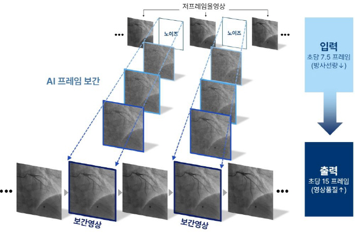
Against this backdrop, Angio-FILM, developed by a research team led by Professor Kang Si-hyuk of the Cardiology Department at Seoul National University Bundang Hospital, with Kwon Hui and Park Se-young as first authors, is a solution that lowers the radiation dose itself while maintaining image quality at the existing level by having AI generate intermediate frames expected to exist between captured images. The system records at about 7.5 frames per second, roughly half the current standard, but uses AI interpolation to fill in missing frames and achieve image quality equivalent to 15 frames per second. The radiation dose that can be reduced through this approach is estimated to be more than half.
The research team said, "Because this is imaging used in precise procedures, it was designed to stably reproduce the heart and coronary arteries' fast and nonlinear motion." They explained that instead of simply deriving values between the preceding and following frames, they improved stability by separating spatial and temporal analysis algorithms and applying a 'Latent Flow Matching' technique that extracts only the key elements of the image and computes the path.
In a Turing test, the team asked 30 specialists to distinguish between original and AI-interpolated versions of 600 videos. Even when they were clearly aware of AI involvement, the probability of identifying the AI-interpolated videos was not significantly different from random selection at 50%. This suggests that the images were rendered with such precision that even specialists could not tell them apart if they tried. The error in coronary lumen diameter between the original and AI-generated images was only 0.18 mm, easing concerns about anatomical distortion.
The study is significant in that it offers a solution capable of producing clinically usable images while reducing radiation exposure in coronary angiography by more than half. Coronary artery disease, which is directly linked to life, is one of the core areas of so-called essential medical care that has recently faced difficulties. Against this backdrop, AI is being credited with opening a breakthrough that could help protect patients and medical staff from radiation exposure.
Professor Kang said, "Physical equipment improvements to reduce radiation dose in coronary angiography have already reached their limits." He added, "Now that this study has secured the clinical reliability of Angio-FILM, its introduction in the field could make a significant contribution to dramatically reducing radiation exposure for patients and medical staff."
Meanwhile, the findings were published in the latest issue of npj Digital Medicine, a sister journal of Nature.